
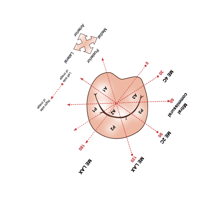
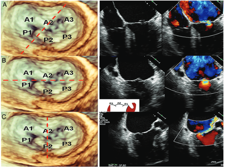
Key TEE views for the assessment of mitral valve morphology: The key views for assessing the mitral valve are demonstrated in these images of a patient with a small P2 prolapse. A 3D TEE en-face view of the MV including segment nomenclature is shown on the left side (A, B, C). The red incised lines illustrate the plane of the corresponding 2D planes on the right side. A mid-esophageal four-chamber view at 0 in (A), an inter-commissural view at 60 [slightly rotated posteriorly in order to better visualize the PISA (proximal isovelocity surface area) of the MR Jet] in (B), and a LVOT view at 150 in (C). The prolapse can be identified in each plane. N. C. Wunderlich and R. J. Siegel. European Heart Journal – Cardiovascular Imaging (2013)14, 935–949doi:10.1093/ehjci/jet060
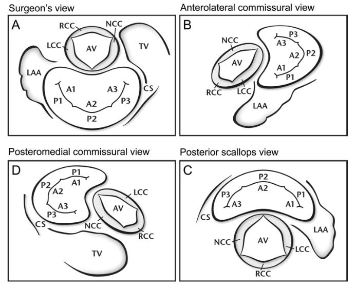
Overview of 4 End-Systolic Standard Views for Assessment of Mitral Valve Prolapse: Each view is oriented about 90° counterclockwise to the previous view. (A) Surgeon’s view: mitral valve “en face” with the aortic valve at 12 o’clock. (B) Anterolateral commissural (CAL) view: obtained by 90 to 110° counterclockwise rotation and 60 to 70° backward tilt of the surgeon’s view. The CAL view is best for evaluation of involvement of the anterolateral commissure as well as A1 and P1. (C) Posterior scallops (PS) view: obtained by 70 to 90° counterclockwise rotation and 20 to 30° medial tilt of the CAL view. The PS view best shows the 3 scallops of the posterior leaflet. (D) Posteromedial commissural (CPM) view: obtained by 90° counterclockwise rotation and slight forward tilt of the PS view. This view is optimal for assessment of the posteromedial commissure, P3 and A3. A1 = segment 1 of the anterior mitral valve leaflet; A2 = segment 2 of the anterior mitral valve leaflet; A3 = segment 3 of the anterior mitral valve leaflet; AV = aortic valve; CS = coronary sinus; LAA = left atrial appendage; LCC = left-coronary cusp of the aortic valve; NCC = noncoronary cusp of the aortic valve; P1= segment 1 of the posterior mitral valve leaflet; P2 = segment 2 of the posterior mitral valve leaflet; P3 = segment 3 of the posterior mitral valve leaflet; RCC = right-coronary cusp of the aortic valve; TV = tricuspid valve.
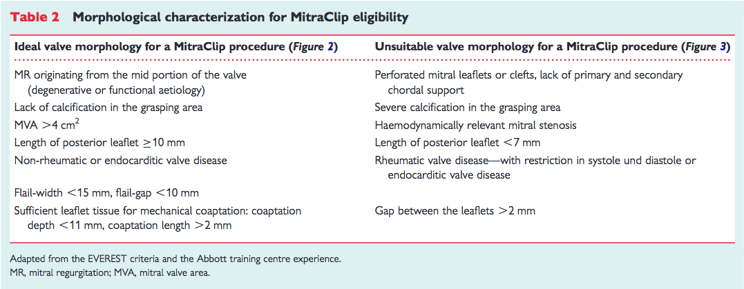

