2D echo:
1. Early diastolic RV collapse
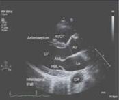
2. RA collapse in atrial diastole (or ventricular systole)
3. LA collapse in atrial diastole
4. Dilated IVC with loss of respiratory collapse
5. Ventricular interdependence
6. Swinging heart
7. LV “pseudohypertrophy” (LV wall thickening that resolves following pericardiocentesis and is hypothesized to be due to myocardial venous congestion in the setting of increased pericardial pressure)
Respiratory variation in Doppler flow:
1. Inspiratory decrease in mitral inflow E-wave velocity >25%.
2. Expiratory decrease in tricuspid inflow E-wave velocity >40%.
3. Hepatic veins: expiratory increase in diastolic flow reversal.
4. Superior vena cava forward flow is predominantly systolic, with decrease or loss of the diastolic component and increased flow reversals on expiration.
5. Left-sided isovolumetric relaxation time is increased in inspiration.
6. Pulmonary veins: inspiratory decrease in velocity.
References:
Questions, Tricks, and Tips for the Echocardiography Boards 1st Edition by Dr. Vincent L. Sorrell MD and Dr. Sasanka Jayasuriya MBBS
Segni ED, Beker B, Arbel Y, et al. Left ventricular pseudohypertrophy in pericardial effusion as a sign of cardiac tamponade. Am J Cardiol 1990;66(4):508–511.
Horowitz MS, Schulz CS, Stinson EB, et al. Sensitivity and specificity of echocardiographic diagnosis of pericardial effusion. Circulation 1974;50:239–47.
Maisch B, Seferović PM, Ristić AD, et al.; Task Force on the Diagnosis and Management of Pericardial Diseases of the European Society of Cardiology. Guidelines on the diagnosis and management of pericardial diseases executive summary; The Task force on the diagnosis and management of pericardial diseases of the European society of cardiology. Eur Heart J 2004;25(7):587–610.